- Home
- Anatomy Guide
- Foot Tendons
Foot & Ankle Tendons
Written By: Chloe Wilson BSc(Hons) Physiotherapy
Reviewed By: FPE Medical Review Board
Damage to the foot and ankle tendons are a common cause of foot pain, typically caused by overuse, overstretching or an injury.
Tendons are thick bands of tissue that connect muscles to bone. When a muscle contracts, the tendon pulls on the bone causing the joint to move.
There are a number of tendons located in the foot and ankle all responsible for different ankle, foot and toe movements. Tendons also help to provide stability around the foot and ankle
Here we will look at where the different foot and ankle tendons are found, what movements they allow, why we need them, what can go wrong and how to treat ankle and foot tendon pain.
Lateral Ankle Tendons
Let’s start by looking at the lateral ankle tendons found on the outer side of the ankle and foot, the peroneal tendons.
1. Peroneal Tendons
There are two peroneal tendons, one from the peroneal longus muscle the other from peroneal brevis.
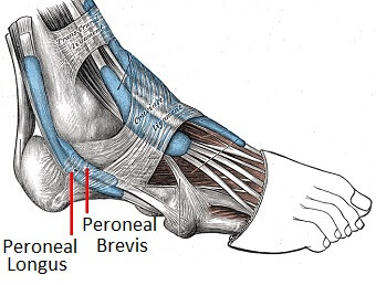
The peroneal tendons run down together behind the outer side of the ankle and then split before attaching to different parts of the foot
Peroneus Longus: Originates from the upper part of the fibula, passes underneath the foot and attaches by the medial foot arch
Peroneus Brevis: Originates from the lower part of the fibula and attaches to the outer side of the midfoot
The peroneal tendons and their respective muscles help to pull the foot down into plantarflexion and outwards into eversion. Functionally, they are very important for providing stability when running, particularly on uneven ground.
RELATED ARTICLE: Peroneal Tendonitis - Causes, Symptoms & Treatment
Medial Foot Tendons
There are a number of foot and ankle tendons that pass around the inner side of the ankle producing various foot movements.
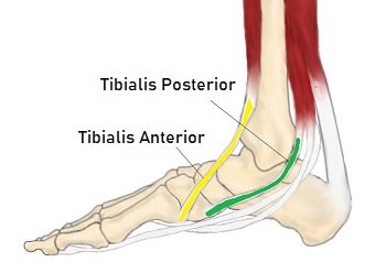
1. Tibialis Anterior Tendon
The tibialis anterior muscle originates from the outer side of the tibia and passes down the front of the shin.
The muscle turns into tendon about two thirds of the way down the shin and travels across the front of the ankle joint to the inner side of the foot underneath the medial foot arch.
Tibialis anterior is a strong ankle tendon that pulls the foot up into dorsiflexion. Functionally, it is really important when walking as it lifts the foot up to prevent it catching on the ground as the leg swings forwards and controls foot placement once the heel strikes the ground.
It also works with other medial ankle tendons to turn the foot inwards into inversion.
RELATED ARTICLE: Tibialis Anterior Tendonitis - Causes, Symptoms & Treatment
2. Tibialis Posterior Tendon
Tibialis posterior is the deepest muscle on the back of the leg. The tendon passes behind the inner ankle bone (medial malleolus) and underneath the foot attaching to the tarsal bones.
The tibialis posterior tendon is the main invertor of the foot and also helps the calf muscles to plantarflex the foot. It plays an important role in supporting the medial arch and functionally controls the position of the foot during walking and running.
RELATED ARTICLE: Posterior Tibial Tendonitis - Causes, Symptoms & Treatment
Posterior Ankle Tendons
A number of muscles and tendons pass down the back of the ankle, but the main one is the Achilles Tendon, aka Calcaneal Tendon.
1. Achilles Tendon
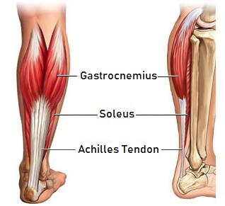
The two main calf muscles, gastrocnemius and soleus, run down the back of the calf and join together to form a strong, thick tendon, the Achilles tendon, that attaches to the back of the heel.
The calf muscles, through the Achilles tendon, are the main plantarflexors of the ankle which pulls the foot down. Functionally, the Achilles ankle tendon provides the large propulsive force needed for walking, running and jumping.
The Achilles tendon is the strongest and thickest tendon in the body and can withstand strains of up to 10 tons.
RELATED ARTICLE: Achilles Tendonitis - Causes, Symptoms & Treatment
Extensor Tendons
There are two sets of extensor tendons found in the foot from three muscles, extensor digitorum tendons and hallucis longus.
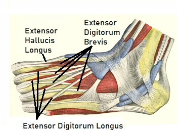
1. Extensor Digitorum Longus: Originates just below the knee on the outer shin bones and forms a tendon before passing across the front of the ankle after which it splits into four foot tendons, one to each of the outer four toes
2. Extensor Digitorum Brevis: Originates from the top part of the heel bone, travels across the top of the foot and has short tendons that attach to the base of the big toe and tips of the second, third and fourth toes.
3. Extensor Hallucis Longus: Originates from part way down the outer shin, passes medially across the front of the shin forming a tendon as it crosses in front of the ankle to the tip of the big toe.
All three of these extensor tendons of the foot work to pull the toes up into extension. Functionally, they raise the toes clear of the ground during running and walking.
RELATED ARTICLE: Extensor Tendonitis: Causes, Symptoms & Treatment
Flexor Tendons
There are two sets of flexor foot tendons, each made up of two muscles and tendons, one set that bends the big toe, the other set that bends the remaining four toes.
1. Flexor Digitorum Brevis: Originates from the bottom of the heel bone and passes underneath the foot to the middle of the sole where it splits into four foot tendons which attach to the base of the outer four toes. The tendons attach to the mid toe rather than extending to the tips.
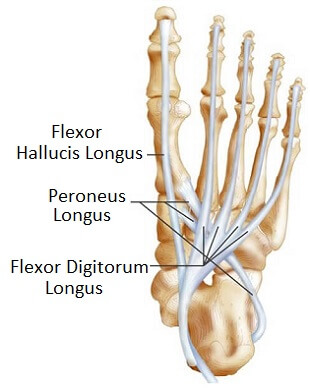
2. Flexor Digitorum Longus: Originates part way down the back of the calf, underneath gastrocnemius and soleus, forming a tendon just above the medial malleolus. It passes behind the inner ankle bone and crosses underneath the sole of the foot where it divides into four tendons that attach to the ends of the outer four toes.
3. Flexor Hallucis Longus: Originates from the back of the lower fibula and crosses medially, where it forms a tendon that passes down the back of the ankle, hooking underneath the inner ankle bones. Continues underneath the foot attaching to the base of the end bone of the big toe, the distal phalanx
4. Flexor Hallucis Brevis: A short muscle that originates deep in the sole of the foot and splits in two before forming short tendons that attach to the base of the first big toe bone, the proximal phalanx
The flexor digitorum tendons and muscles bend the outer four toes. Functionally, they pull the toes down into the ground allowing the foot to both grip the floor for balance and thrust off through the toes when walking, running and jumping.
The flexor hallucis tendons and muscles bend the big toe down, and the longus muscle also helps the calf muscles to plantarflex the foot, and helps support the foot arches. Functionally, these big toe flexor tendons are really important as they produce the final thrust from the foot when walking.
Foot & Ankle Tendon Injuries
The foot and ankle tendons are usually damaged by
- Repetitive Overloading: Excess force repeatedly placed through the tendon e.g. sports leads to inflammation and degeneration
- Friction: Repetitive pressure rubbing on the tendon e.g. tight footwear – also results in inflammation and degeneration
- Overstretching: Muscle and tendon stretched beyond their elastic limit resulting in tearing
Foot tendon pain may be due to:
- Tendonitis: When the foot or ankle tendons become inflamed causing pain and swelling LEARN MORE >
- Tendonosis: Wear and tear or degeneration of the foot tendon
- Tendon Tears: Where the foot or ankle tendon partially or completely ruptures

Treating Tendon Injuries
Treatment for foot and ankle tendon injuries will depend on which tendon is damaged and the severity of the injury and foot tendon pain, but typically consists of
- Rest: from any aggravating activities so as not to overload the tendon
- Ice: Regularly applying ice packs can help reduce pain and inflammation with foot tendon injuries LEARN MORE >
- Compression Bandage: helps to support the ankle and foot as well as reduce swelling
- Elevation: helps to reduce swelling
- Immobilisation: A splint or cast may be worn to prevent movement so the tendon can heal. In severe cases crutches may be necessary to keep the weight of the affected foot
- Medication: painkillers and non-steroidal anti-inflammatories can be a big help with foot tendon pain
- Physical Therapy: Friction massage and ultrasound can help with tendon injuries
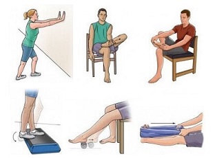
- Exercises: Strengthening and stretching exercises help the foot tendons to heal correctly and reduce the risk of future injury LEARN MORE >
- Orthotics: Shoe inserts can be helpful for offloading tendons and ensuring the foot is in the correct position so as not to place excess strain on the foot tendons LEARN MORE >
- Injections: Some ankle tendon injuries respond well to steroid injections which reduce pain and inflammation LEARN MORE >
- Surgery: In some instances injury to the foot or ankle tendons may require surgery. This is usually because either the tendon has been ruptured completely and thus needs to be sewn back together, or when symptoms fail to settle after six months of ongoing treatment
What Is Causing My Foot Tendon Pain?
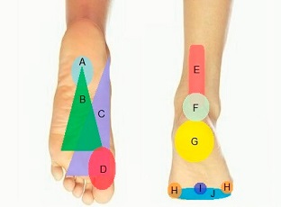
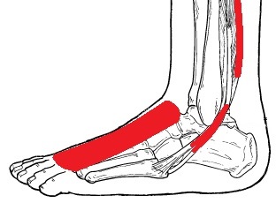
Foot and ankle pain is often due to tendon injuries. The location of the pain will depend on which tendon is damaged
- Outer Foot/Ankle Pain: Peroneal Tendon Injury
- Inner Foot/Ankle Pain: Posterior Tibial Tendon Injury
- Heel/Calf Pain: Achilles Tendon Injury
- Top of Foot Pain: Extensor Tendon Injury
- Front Of Ankle Pain: Tibialis Anterior Tendon Injury
You can find out loads more about each of these causes of foot tendon pain, including symptoms, diagnosis and treatment ideas by using the links above. Alternatively, visit the Foot Tendonitis page.
The foot and ankle tendons play a really important role in day to day life, so any problems deserve prompt treatment.
You may also be interested in the following articles:
Related Articles
Page Last Updated: 05/04/23
Next Review Due: 05/04/25
